Why It Happens and How It Can Be Corrected
One of the most recognizable complications after lower eyelid surgery is inferior scleral show. Patients usually notice that the lower eyelid sits too low beneath the eye, exposing the white of the eye below the iris. The eye may appear rounder than before surgery or simply different.
Some patients first notice the changein photographs. Others feel the problem before they fully see it. Their eyes feel dry, irritated, or exposed. Contact lenses become difficult to wear. The eyelids may feel tight or uncomfortable.
Inferior scleral show is not simply a cosmetic issue. It is often a sign that the lower eyelid support system has been disrupted.
In most cases following blepharoplasty, inferior scleral show reflects lower eyelid retraction.
What Inferior Scleral Show Means
In a natural eyelid position, the lower eyelid margin rests at or very close to the lower edge of the iris. When the eyelid drops lower than this position, the white sclera becomes visible beneath the iris.
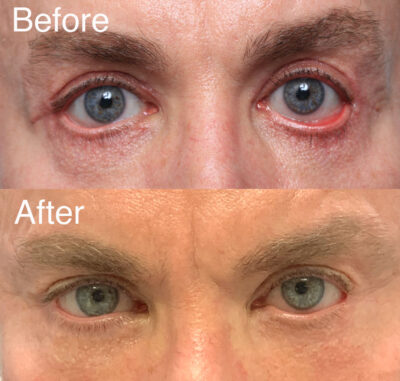
This visible band of white is what surgeons call inferior scleral show.
Patients often describe it in simpler terms:
• “My eye looks too open.”
• “The lower lid looks pulled down.”
• “The eye looks round instead of almond shaped.”
Those descriptions are accurate. The normal almond contour of the eye depends heavily on proper lower eyelid position.
When the lower eyelid drops, the eye takes on a more rounded appearance.
Why Scleral Show Happens After Blepharoplasty
Lower eyelid surgery changes the anatomy of the eyelid in several ways. When everything heals properly, the eyelid looks smoother and more youthful.
When things do not go as planned, several structural problems may develop.
Loss of vertical eyelid height
Lower eyelid surgery often involves removal of skin or muscle. If too much tissue is removed, the eyelid becomes shorter vertically.
During healing, scar contraction can pull the eyelid downward even further.
Weakening of the orbicularis muscle
The orbicularis oculi muscle acts like a muscular hammock supporting the eyelid margin. Incisions beneath the eyelashes can weaken that support system.
Some evidence suggests that branches of the facial nerve supplying the muscle may also be affected during surgery. When this muscular support is compromised, the eyelid margin becomes less stable.
Scar formation within the eyelid
Scar tissue can develop within multiple layers of the eyelid after surgery. These scars can restrict movement and contribute to downward pull on the eyelid.
Lack of support from the cheek and orbital rim
One of the most overlooked contributors to lower eyelid retraction is the relationship between the eye and the bone beneath it.
The lower eyelid normally rests on the cheek. If the cheek lacks adequate projection or support from the orbital rim, the eyelid is left vulnerable to downward pull.
Patients with prominent eyes or a negative vector eyelid are particularly susceptible.
Why Tightening the Eyelid Often Does Not Work
Many patients researching treatment encounter procedures such as:
• lateral canthopexy
• lateral canthoplasty
• drill-hole canthoplasty
These procedures tighten the outer corner of the eyelid.
They can be useful in selected cases where eyelid laxity is the primary issue. Unfortunately, that is rarely the full story after blepharoplasty.
In most cases of inferior scleral show, the real problem is loss of vertical eyelid support, not horizontal laxity.
Simply tightening the eyelid laterally can apply the wrong forces to the eyelid.
A useful analogy is tightening a belt to hold up pants resting on a protruding abdomen. The belt becomes tighter, but the pants may slide downward instead of staying up.
In the same way, tightening the outer corner of the eyelid may pull the shortened eyelid under the curvature of the eye rather than restoring normal height.
Some patients undergo multiple procedures of this type without solving the underlying problem.
A Better Way to Think About Lower Eyelid Retraction
When I evaluate patients with inferior scleral show after blepharoplasty, I look at several structural issues.
Has the eyelid been shortened vertically?
Has scar contraction stiffened the eyelid?
Has the muscular support system been weakened?
Does the cheek provide enough support for the eyelid?
Does the orbital rim have enough projection to support the midface?
These questions help determine what kind of reconstruction will work.
The key concept is simple: the lower eyelid is supported from below. If that support system is not restored, tightening the eyelid alone will not solve the problem.
The Importance of Midface Support
The lower eyelid rests on the cheek. When the cheek lacks structural support, the eyelid pays the price.
One of the major advances in treating lower eyelid retraction has been recognizing the role of the midface and orbital rim.
In my approach, I use a vertical midface lift combined with a hand-carved ePTFE orbital rim implant.
The implant restores projection along the orbital rim and provides a strong surface to which the lifted cheek tissues can be secured.
This allows the cheek soft tissue to be recruited vertically and permanently anchored at the orbital rim.
Instead of forcing the eyelid upward under tension, the eyelid gains support from below.
The Role of the Hard Palate Graft
Even when cheek support is restored, the eyelid itself often requires reconstruction.
For this purpose I commonly use a hard palate graft harvested from the roof of the mouth.
This graft acts as a structural spacer within the lower eyelid. It restores vertical dimension and stabilizes the eyelid margin.
Hard palate tissue works well for this purpose because it is:
• firm and durable
• biologically compatible
• resistant to shrinkage
Because the graft comes from the patient’s own tissue, there is no risk of rejection and minimal inflammation compared with some other materials.
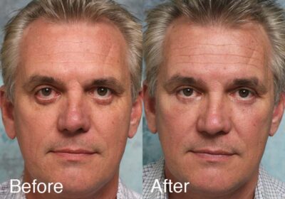
Restoring the Natural Shape of the Eye
When the eyelid and midface support system are reconstructed properly, the lateral corner of the eye can be restored with minimal tension.
This is important.
The goal is not simply to pull the eyelid upward. The goal is to rebuild the anatomy so the eyelid sits naturally against the eye.
Patients often notice several changes after reconstruction:
• the eye looks less round
• the eyelid contour looks more natural
• dryness and irritation improve
• the midface appears more youthful
The improvement in facial balance is often a welcome additional benefit.
Why Revision Surgery Requires Experience
Revision lower eyelid surgery is complex. Each prior procedure may consume valuable tissue or alter anatomy in ways that make reconstruction more difficult.
A key question in revision surgery is simple:
How will the next surgery be different from the ones that failed?
Successful reconstruction requires restoring structure, not simply repeating a tightening procedure.
About Dr. Steinsapir
Dr. Steinsapir is a board-certified eye surgeon and fellowship-trained in oculoplastic surgery and cosmetic surgery in Beverly Hills, California where he specializes in balanced facial cosmetic surgery for natural results, with an emphasis on minimally invasive techniques, fast recovery time, and leadership in medical technology. Dr. Steinsapir has a private practice and also serves as an Attending Surgeon at UCLA/Harbor Medical Center. He is on staff at the David Geffen School of Medicine at UCLA. Contact us today to learn how Dr. Steinsapir’s experience and training make him an expert in cosmetic surgery, which can be a vital part of your evidence-based treatment plan.
Services described may be “off-label” and lack FDA approval. This article is informational and does not constitute an advertisement for off-label treatment. No services should be provided without a good faith examination by a licensed physician or surgeon and an informed consent with a discussion of risks, benefits, alternatives, and the likelihood of treatment success. Only you and your treating physician or surgeon can determine if a treatment is right for you.
Frequently Asked Questions About Inferior Scleral Show
What is inferior scleral show?
Inferior scleral show refers to the white of the eye becoming visible beneath the iris because the lower eyelid sits too low.
Is scleral show after blepharoplasty common?
It is an uncommon but well-recognized complication of lower eyelid surgery. It is most often associated with transcutaneous lower blepharoplasty.
Does scleral show always mean something went wrong with surgery?
Not always. Some patients naturally show a small amount of sclera beneath the iris. Some individuals have a large eye relative to orbit size. When this primarily is visible at the orbital rim, which is common, a midface lift with rim implant and hard palate graft is an excellent and safer alterative to orbital decompression surgery. New or worsening scleral show after surgery usually indicates lower eyelid retraction caused by surgery.
Can scleral show improve on its own?
Very mild cases may improve as swelling resolves. True lower eyelid retraction caused by structural shortening usually does not correct itself.
Can fillers help treat scleral show?
Hyaluronic acid filler can sometimes improve mild cases by stiffening the eyelid and supporting the tissues. The effect is temporary and does not correct structural eyelid shortening.
Why doesn’t canthoplasty fix the problem?
Because canthoplasty primarily tightens the eyelid horizontally. Most cases of post-blepharoplasty scleral show involve loss of vertical support.
What is the most reliable surgical treatment?
In many cases, durable correction requires restoring support beneath the eyelid and rebuilding the eyelid itself. This may involve midface lifting, orbital rim augmentation, and spacer graft reconstruction.
What is a hard palate graft?
A hard palate graft is tissue taken from the roof of the mouth and used to stabilize and lengthen the lower eyelid. It is a well-established technique in eyelid reconstruction.
Is the donor site painful?
The palate donor site is sore for several days but generally heals quickly. An acrylic palate stent that looks like a retainer substantially improves comfort and virtually eliminates the small risk of bleeding.
Why is an in-person evaluation important?
Lower eyelid retraction involves multiple anatomical factors that cannot be fully evaluated through photographs or video calls. A detailed physical examination is essential to determine the proper treatment. Don’t get talked into a video chat as your surgical consultation. Yes, there are surgeons who exactly practice this way. You don’t actually meet them in person until the day of surgery. Ask yourself if that type of approach makes any sense? We don’t think so.