310.274.7422
9001 Wilshire Blvd, Suite 305, Beverly Hills, CA 90211

Revision

3141

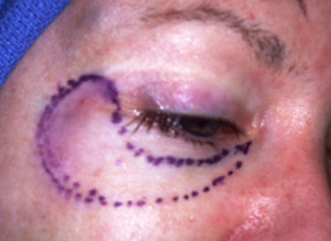
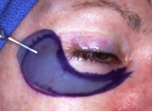
Note this case contains graphic images from surgery.

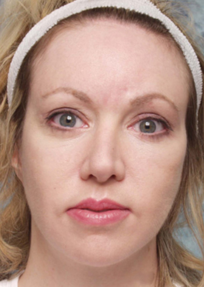
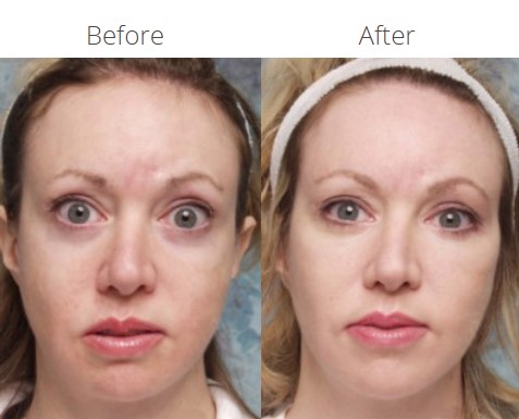
Cosmetic Concern: 34-year old woman with a history of craniofacial surgery for maxillary hypoplasia. To address remaining aesthetic concerns she also had eyelid surgery and several facelifts. However, she feels that she still lacks the projection of her cheekbones that would be best for her face.

Diagnosis:

  1. Glabellar scar secondary to flap perforation during forehead surgery.
  2. Bilateral upper eyelid retraction.
  3. Maxillary hypoplasia with deficient inferior orbital rim. Projection.
  4. Marked facial asymmetry.

Treatment:

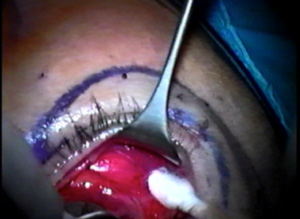
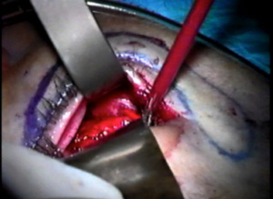
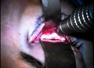
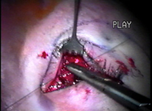
  1. Augmentation of the orbital rims with custom hand carved orbital rim implant made from ePTFE.
  2. Vertical midface lift.
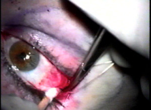
  3. Bilateral lateral canthoplasty

Discussion: The patient needed additional volume in the midface. The ePTFE rim implant is custom carved for her particular anatomic deficit. (Figures A-C)

fig-a
Figure A. Area needing augmentation.


Figure B. A template is fabricated and transferred to the sheet of ePTFE.
fig-b


Figure C. Implant material is hand shaped to the desired form.
fig-c


The midface is expose using a tiny lateral canthal incision. (Figure D).
fig-d
Figure D. A lateral canthotomy and cantholysis is performed.


A dissection is carried out to expose the orbital rim and the bone of the midface. (Figure E).
fig-e
Figure E. Dissection is carried out behind the eyelid to expose the orbital rim.


A periosteotomy is performed which cuts the dense connective tissue that covers the bone. (Figure F)
fig-f
Figure F. After degloving the midface skeleton, a periosteotomy is performed to mobilize the cheek.


This allows the cheek soft tissue to be mobilized and vertically elevated. The implant is positioned on the orbital rim. (Figure G)
fig-g
Figure G. The hand carved ePTFE implant is positioned to sit along the orbital rim.


The implant is fixed to the top of the orbital rim with titanium microscrews so the implant is immobilized. Figure H)
fig-h
Figure H. Holes are drilled for the placement of titanium microscrews, which will permanently hold the implant in position.


Stitches are then used to lift the cheek soft tissue to the top of the ePTFE orbital rim implant. (Figure I)
fig-i
Figure I. Sutures from the top of the implant are used to vertically lift the cheek soft tissues.


Finally, the cut edge of the lower eyelid is resuspended to the orbital rim using a procedure known as a canthoplasty. (Figure J)
fig-j
Figure J. Placement of the deep supporting stitch that will hold the lateral eyelid tissue to the orbital rim at just the right position is a critical step in the cathoplasty.

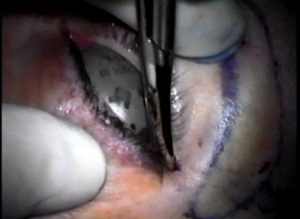
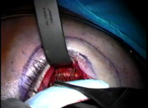
The outer aspect of the upper eyelids are reattached to the newly formed lateral canthal angle correcting the upper eyelid retraction. Finally, the skin is closed completing surgery.

Before
Revision
After
Revision