Loading gallery items...
Reconstruction
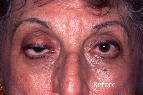
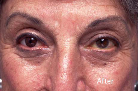
- Notes: 75-year old woman lost the use of her right eye as a child. She wears a glass shell over the right eye. For the past 50 years, her right upper eyelid has been droopy. Diagnosis: Non-seeing right eye. Right upper eyelid ptosis Treatment: Right upper eyelid ptosis surgery. Hand painted contact lens for the right eye. Discussion: Once the right upper eyelid ptosis surgery was performed, the ocularist created a hand painted contact lens completing the rehabilitation.
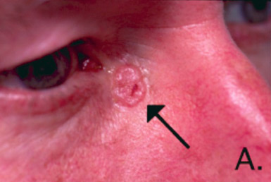
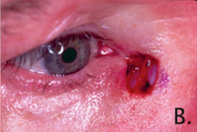
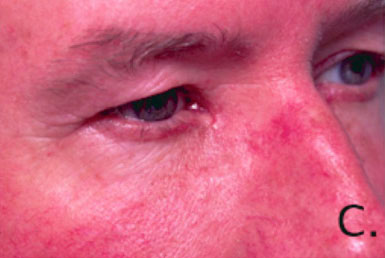
- Notes: Dr. Steinsapir provides comprehensive management for eyelid skin cancers. His expertise is widely recognized. The most common skin cancers of the eyelid are basal cell carcinoma and squamous cell carcinoma. These are usually caused by long-term sun exposure. Dr. Steinsapir typically works with a Mohs’ cancer surgeon who performs the excision of the lesion using microscopic control. Dr. Steinsapir then does the reconstruction. This team approach provides the highest rate of cure and at the same time conserves the uninvolved eyelid by limiting the removal of normal tissue. It is Dr. Steinsapir’s goal to provide the most natural reconstruction possible.
- Notes: 74-year old with long standing left facial paralysis following the removal of an acoustic neuroma from the brain stem. Diagnosis: Left facial paralysis. Marked left lower eyelid atrophy. Left lower eyelid vertical inadequacy Treatment: Custom ePTFE left orbital rim implant. Left midface lift. Alloderm implant to the left lower eyelid.
- Notes: 26 year-old woman with a life long history of a slowly expanding right orbital rim mass. She had seen physicians in the past and was advised that it was fat and that surgery was unnecessary. Dr. Steinsapir ordered a CT scan to assess the lesion. The arrows on the two CT images point to the lesion.
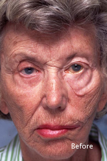
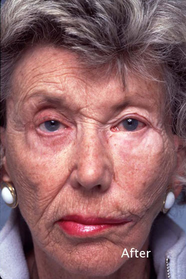
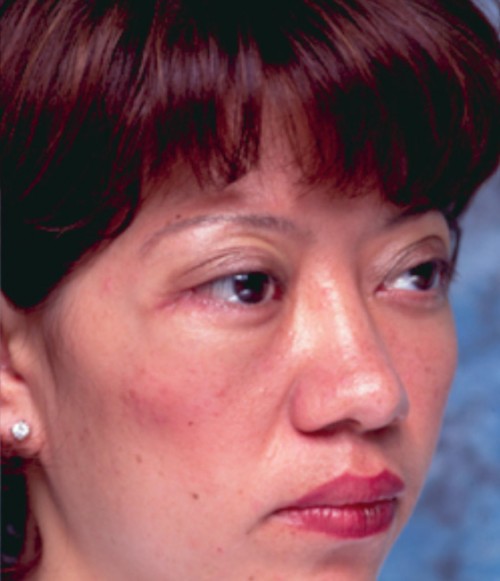
- Notes: This 51-year old woman lost her left eye from a childhood orbital tumor. She consulted Dr. Steinsapir because her glass eye would not stay behind the left eyelids.
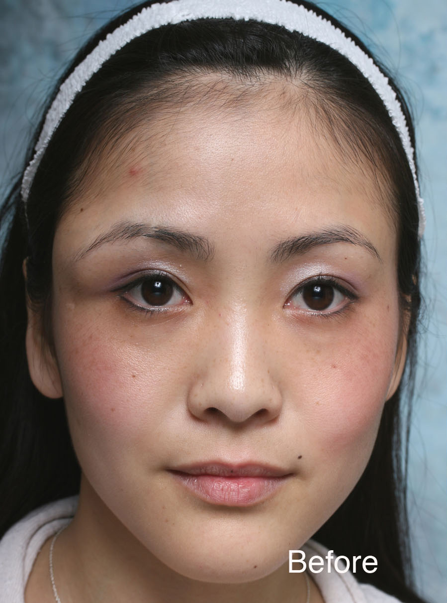
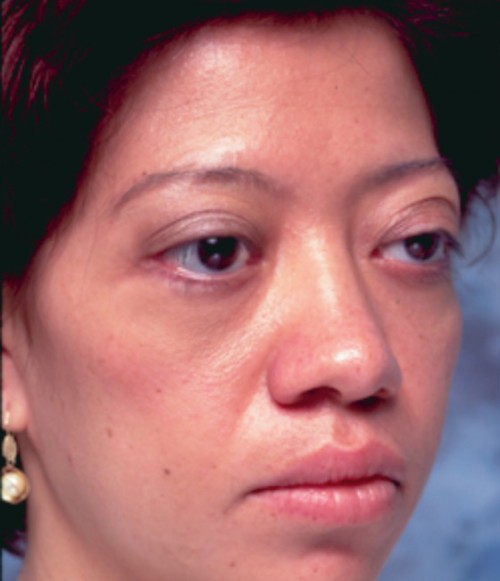
- Notes: This 28-year old has a history of childhood thyroid eye disease. She had orbital decompression at the Jules Stein Eye Institute as well as several other eyelid procedures. However, she has difficulty closing the eyelids.
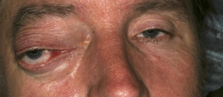
- Notes: This 45 year man (Figure 1) reports that over a period of years he has noticed a gradual swelling the right inner corner of his eyelids with a progressive bulging and displacement of the eye. He is now seeking care because the process seems to be affecting his vision.
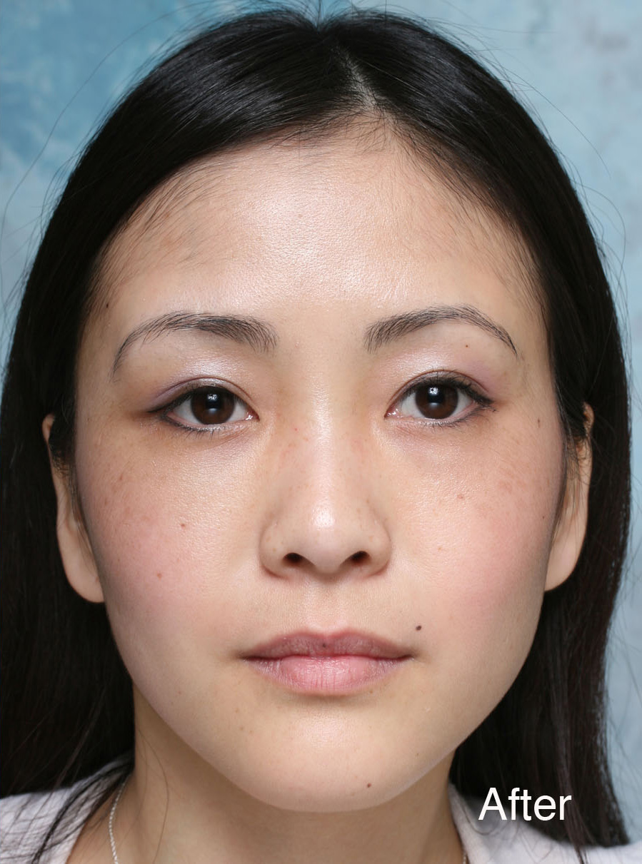
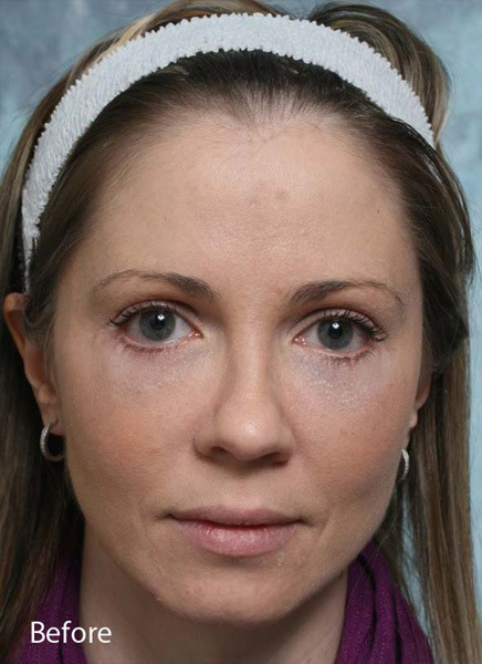
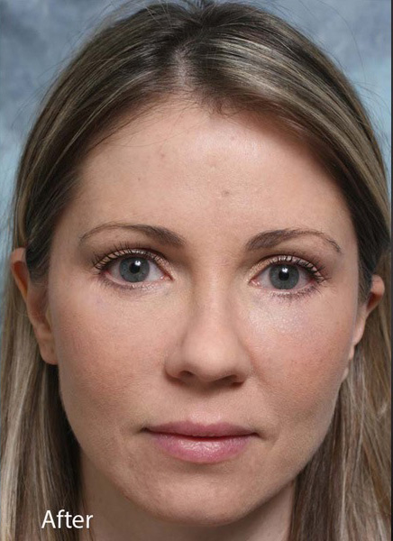
- Notes: 33 year with lower eyelid compromise after lower transcutaneous blepharoplasty. Diagnosis: The lower eyelids have been pulled down do to lack of skin and scaring caused by her original lower eyelid surgery. The lower eyelids eyes are bowed down with inferior scleral show. There is also midface ptosis. Treatment: Dr. Steinsapir has pioneered the use of hand carved orbital rim implants to support the lower eyelid. Also hard palate graft has proven far more reliable for reshaping the lower eyelids than products like Alloderm or Enduragen. She had surgery is two operative sessions and is two months out from the second procedure.
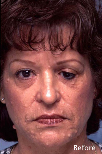
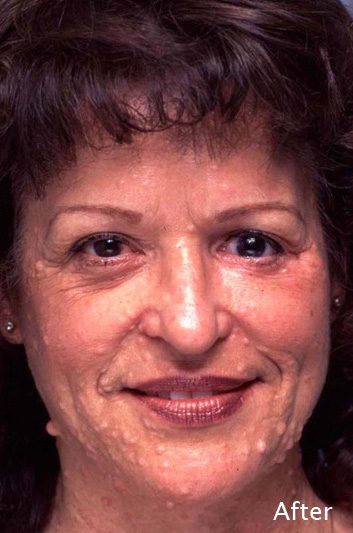
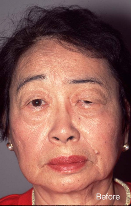
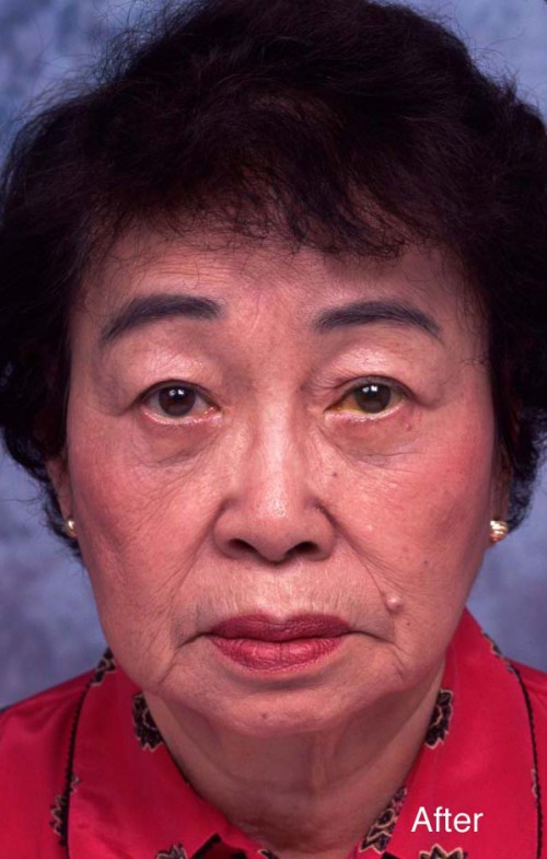
- Notes: 74-year-old woman with left facial weakness following Bell’s palsy. Diagnosis: Bell’s palsy. Left forehead paralysis. Pseudoherniated orbtial fat. Treatment: Endoscopic forehead lift. Upper eyelid surgery. Lower eyelid surgery.
No items match your filters.